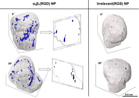
Figure 5.
3D reconstructions of MR signal enhancement reveal tumor neovascular morphology. The tumor volume is outlined in gray, and voxels meeting the enhancement threshold at 2 h postinjection of contrast agent are shown in blue. Left panel: two rotated views of an α5β1(RGD)-targeted tumor. The cross sections at right demonstrate the paucity of angiogenesis in the core. Right panel: minimal enhancement associated with irrelevant RGS-targeted contrast agent.

