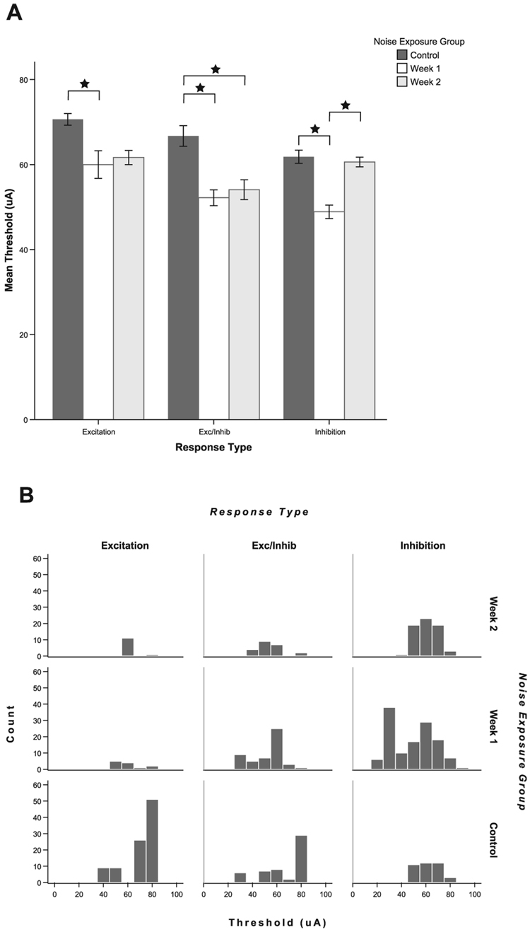
FIG. 9.
Thresholds to trigeminal stimulation are significantly decreased following noise damage, reflecting increased sensitivity to trigeminal stimulation in the deafened animals. (A) Mean thresholds to trigeminal stimulation are decreased at both 1 and 2 weeks after noise exposure. At 1 week following noise exposure, thresholds are significantly lower for all response types [excitatory (E), excitatory/inhibitory (E/In) and inhibitory (In)]; at 2 weeks, thresholds for E and E/In units are significantly lower but In response thresholds have returned to normal. *Significant pair-wise differences after Bonferroni correction (see text). Error bars indicate ± 1 SEM. (B) The frequency distribution plots indicate a sharp peak in the number of low threshold In units at 1 week following noise damage and a reduction in the number of high threshold units for the E and E/In groups after noise damage.

