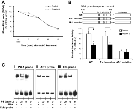
Figure 5.
Effect of human protein S on the regulation of SR-A expression. (A) SR-A mRNA stability. THP-1 macrophages were treated with 5 μg/mL actinomycin D (Act-D) in the presence or absence of 20 μg/mL protein S for 16 hours. Total RNA was harvested at the indicated times and analyzed by real-time PCR using SR-A and 18S rRNA primers. Data are shown as the percentage of mRNA remaining in respect to cells before the addition of Act-D. Representative results from 3 experiments are shown. (B) SR-A promoter activity. THP-1 cells were transiently cotransfected by electroporation with SR-A gene promoter constructs (WT, PU.1 mutation, and AP-1 mutation) and pRL-SV40 plasmids as an internal control. After the transfection, the cells were treated with PMA (50 ng/mL) for 24 hours, followed by protein S (20 μg/mL) treatment for 24 hours. Dual-luciferase assay was performed. Columns and vertical bars denote the mean plus or minus SD of 3 experiments. *P < .05 versus untreated controls. (C) PU.1-, AP-1–, and Ets-binding activities. Nuclear extracts were prepared. Electrophoretic mobility shift assay (EMSA) was performed using biotin end-labeled double-stranded oligonucleotides including PU.1, AP-1, and Ets consensus sequences in the SR-A promoter. The transcription factor–probe mixtures were separated by electrophoresis and transferred to a nylon membrane. The signal was detected with an EMSA kit (Panomics). Representative results were from 3 experiments. (i) PU.1 probe. (ii) AP-1 probe. (iii) Ets probe.

