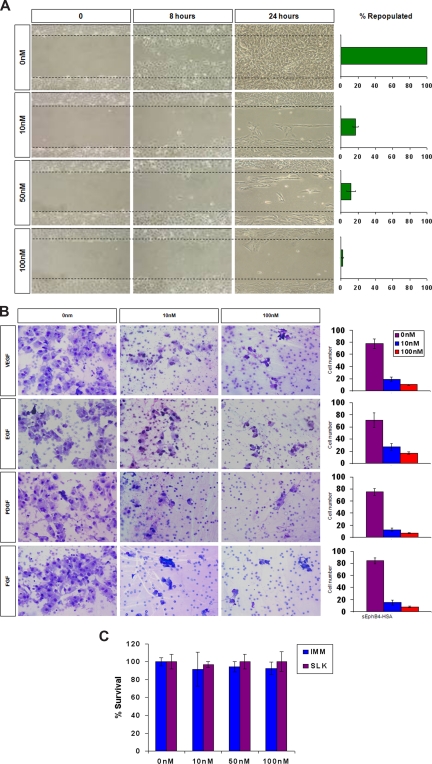
Figure 3.
sEphB4-HSA activity in KS migration, invasion, and survival. (A) KS-SLK cells were grown to confluence, scraped, and treated with varied concentrations of sEphB4-HSA. Cell migration in the clear zone was documented by photographs at various time points at 20× fields. (B) KS cell invasion in response to growth factors. Modified Boyden chamber assay was used to determine KS cell invasion across Matrigel-precoated inserts. Data are presented as number of invading cells plus or minus SE from duplicate wells in 2 experiments. (C) Cell viability assay. KS cells were grown in triplicate in the presence of increasing concentrations of sEphB4-HSA for 72 hours. Cell viability was assessed by MTT assay. The experiment was repeated twice with similar results. Photomicrographs in panels A and B were taken with a Nikon Coolpix 5000 camera (Nikon, Tokyo, Japan) and a Carl Zeiss Invertoskop microscope (Zeiss, Goettingen, Germany) with a 4×/0.12 NA objective and 10× eyepiece.

