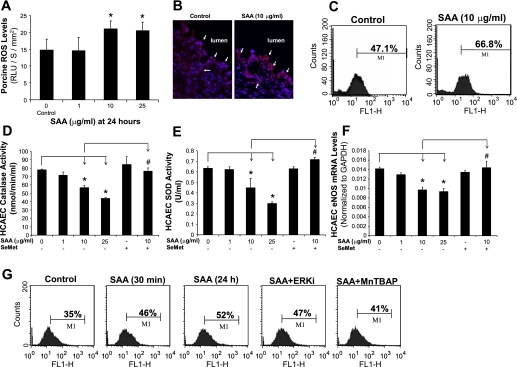
Fig. 5.
Role of SAA-induced oxidative stress on endothelial dysfunction in both porcine coronary arteries and HCAECs. A: ROS levels in the endothelial layer of porcine coronary arteries were tested with lucigenin-enhanced chemiluminescence assay. SAA significantly increased ROS levels of vessel rings in a concentration-dependent manner compared with controls. RLU, relative light units. B: sections of frozen and unfixed porcine coronary artery rings were stained with a fluorescent oxidative dye [dihydroethidium (DHE)] to show ROS levels (red) and counterstained with 4′,6-diamidino-2-phenylindole (blue). ROS staining was increased in SAA-treated vessels in both endothelial and smooth muscle cell layers compared with control vessels. Magnification: ×400. C: ROS levels in HCAECs were stained with DHE and analyzed by FACS Calibur flow cytometry. ROS levels in SAA-treated HCAECs were substantially increased. D and E: activities of both catalase [CAT (D)] and SOD (E) in HCAECs were significantly decreased in SAA-treated cells in a concentration-dependent manner. Coculture with the antioxidant seleno-l-methionine (SeMet) effectively blocked the SAA-induced decrease in activities of both enzymes. F: SeMet effectively prevented the SAA-induced decrease of eNOS mRNA levels in HCAECs. n = 3. G: ROS levels in HCAECs were substantially increased in the early stage of 10 μg/ml SAA treatment (30 min), and the antioxidant Mn(III) tetrakis-(4-benzoic acid)porphyrin [MnTBAP (SOD memetic)] effectively blocked the SAA-induced increase in ROS levels. ERKi, ERK inhibitor. *P < 0.05, controls (DMSO) compared with SAA; #P < 0.05, SAA compared with SeMet + SAA.

