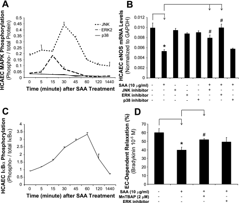
Fig. 6.
Effects of SAA on the phosphorylation of MAPKs and IκB-α in HCAECs. HCAECs were treated with SAA (10 μg/ml) for different times. Phosphorylated and total ERK2, JNK, and p38 as well as IκB-α were detected by Bio-Plex immunoassay. A: SAA (10 μg/ml) treatment increased the ratios of phosphorylated and total ERK1/2 and JNK, but not p38, at 15 and 30 min, respectively. B: to confirm the functional role of these MAPKs in SAA action, JNK inhibitor (SP-600125, 40 μM), ERK1/2 inhibitor (PD-98059, 40 μM), or p38 inhibitor (SB-239036, 1 μM) was used to pretreat HCAECs for 1 h before SAA (10 μg/ml) treatment. eNOS mRNA levels were analyzed with real-time PCR. JNK and ERK1/2 inhibitors, but not the p38 inhibitor, effectively blocked the SAA-induced eNOS decrease. C: SAA (10 μg/ml) treatment induced a gradual increase of IκB-α phosphorylation levels starting from 15 min and peaking at 60 min. n = 3. D: blocking effects of MnTBAP (SOD mimetic) or PD-98059 on the SAA (10 μg/ml)-induced decrease in endothelium-dependent relaxation in response to bradykinin (10−5 M). n = 4. *P < 0.05, controls (DMSO) compared with SAA; #P < 0.05, SAA compared with JNK inhibitor + SAA, ERK1/2 inhibitor + SAA, or MnTBAP + SAA.

