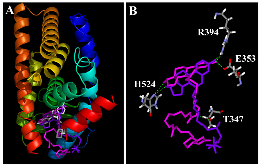
Figure 4.
Interactions of raloxifene (RAL), ICI-182,780 and compound 7 with the ligand binding domain (LBD) of human ERα. A. The three-dimensional structures of ERα LBD in complex with RAL (magenta), ICI-182,780 (purple) and compound 7 (white). The secondary structure of ERα (in ribbon) is colored from blue (N-terminus) to red (C-terminus). This figure is drawn using the PyMOL software. B. Key amino acid residues in ERα LBD that interact with ICI-182,780 and compound 7. Amino acids are colored for their elements (white for hydrogen, red for oxygen, and blue for nitrogen). Compound 7 is colored in magenta and ICI-182,780 in purple. The green dashes show hydrogen bonds between ligands and ERα. This figure is drawn using the DiscoveryStudio software.

