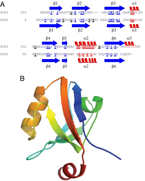
Fig. 1.
Sequence and structure of PSD-95 PDZ3. (A) Sequence alignment of PSD-95 PDZ3 and PTP-BL PDZ2. The secondary structure in the native state is also shown. Positions that were mutated in this study and in ref. 34 are underlined and in bold. (B) Native structure of PSD-95 PDZ3, rainbow colored from the N terminus (blue) to the C terminus (red).

