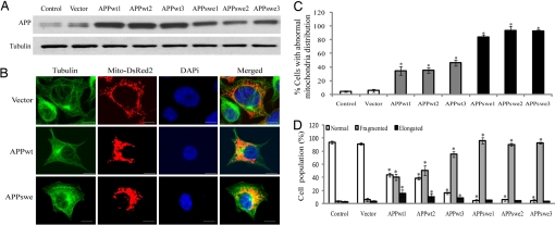
Fig. 1.
APP causes abnormal mitochondrial dynamics in M17 cells. (A) Representative immunoblot of APP. Tubulin is used as an internal loading control. (B) Representative pictures of stable M17 lines with different mitochondrial distribution patterns are shown. Green, tubulin; red, mito-DsRed2; blue, DAPi. Mitochondrial distribution (C) and morphology (D) are analyzed. At least 1,000 cells were measured for each cell line. (Scale bars, 10 μm.) *, P < 0.05, Student t test.

