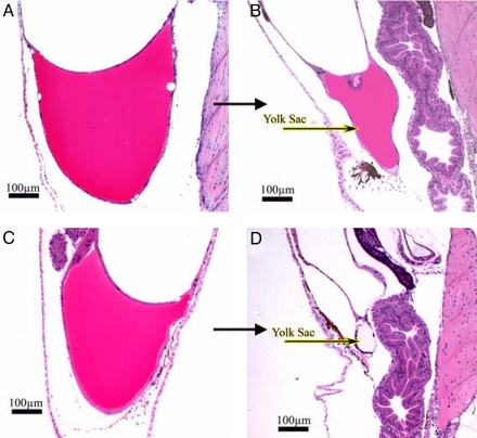
Fig. 4.
Histopathological evaluations of yolk sacs of river and hatchery larvae. Striped bass larvae from hatchery females (n = 15) and 3–5 river-collected females (n = 75) were embedded in glycol methacrylate and serial sectioned at 4-μm thickness by using a Sorval JB-4A Microtome. Sections were placed onto coded and numbered glass slides, stained with H&E, and coverslipped by using Shandon-Mount. (A and C) Yolk sac appearance on day 3 of larvae from hatchery striped bass females (A) and river-collected females' larvae (C). (B and D) Yolk sac appearance on day 5 of larvae from hatchery striped bass females (B), and depleted yolk sac from river-collected females' larvae (D).

