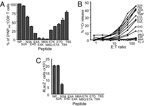
Fig. 1.
Residues important for TCR recognition by DbNP366+CD8+ T cells. (A and B) Splenocytes obtained from PR (H1N1)-primed mice secondarily-infected with the WT HK (H3N2) influenza virus were stimulated with either the WT NP366–374 or a panel of mutant peptides with single amino acid substitutions. The extent of TCR recognition was assessed by IFN-γ production in the ICS assay (A) and 51Cr cytotoxicity after incubation with target EL-4 cells pulsed with 1 μM peptides and 750 μCi 51Cr (B). (C) The response profiles of monoclonal DbNP366+ LacZ-inducible hybridomas expressing the public SGGGNTGQL CDR3β (11) to WT NP366 and NP-mutant peptides are shown.

