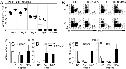
Fig. 2.
Virus clearance and CD8+ T cell profiles after infection with the WT HK and mutant HK-NPM6A viruses. Mice were infected with WT HK or the mutant HK-NPM6A virus. (A) Lungs were sampled at days 3, 6, 7, 8, and 9 after infection and titrated in a plaque assay. The results are log10 pfu per lung (n = 5). (B) CD8+ T cell responses were measured after 10 (day 10) or 20 (day 8) i.n. challenge of naïve or PR- or PR-NPM6A-primed mice with the homologous HK WT of HK-NPM6A virus. Cells were stained with the DbNP366-APC, DbNP-M6A-APC, or DbPA-PE tetramers.(C–F) The response magnitude was assessed for spleen (C and E) and BAL (D and F) sets by ICS. Datasets are mean ± SD for n = 5.

