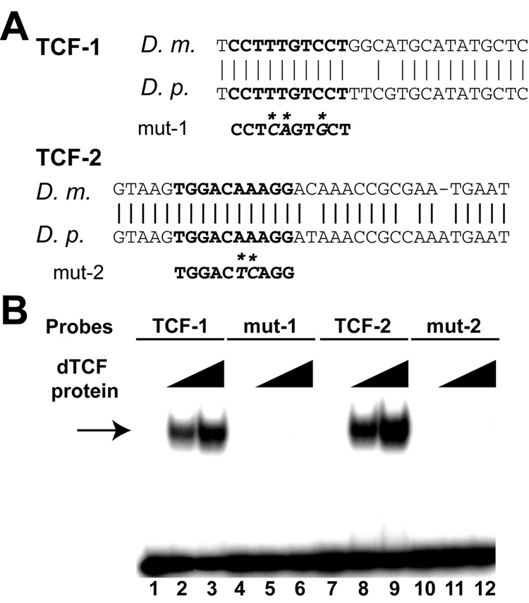
Fig. 5. Two conserved TCF-binding sites in the 4.6-PNS enhancer.
(A) Alignment of sequences around the conserved TCF-binding sites between D. melanogaster and D. pseudoobscura. The bold letters represent the TCF-binding sites in the 4.6-PNS enhancer. The sequences of the mutant TCF sites are shown below the binding sites and base pair changes are indicated by “*”. (B) Gel shift assay showing that the Drosophila TCF protein bound strongly and specifically to the wild type TCF1 (lanes 2 and 3) and TCF-2 (lanes 8, 9). In contrast, no binding was observed when the TCF sites were mutated (mut-1 and mut-2, lanes 5, 6 and 11, 12). No dTCF protein were added in lanes 1, 4, 7, and 10; half the amount of dTCF protein was added to lanes 2, 5, 8, 11 compared to lanes 3, 6, 9, and 12. An arrow points to the dTCF-DNA complex. The sequences of TCF and mutated TCF probes are shown in Materials and Methods.

