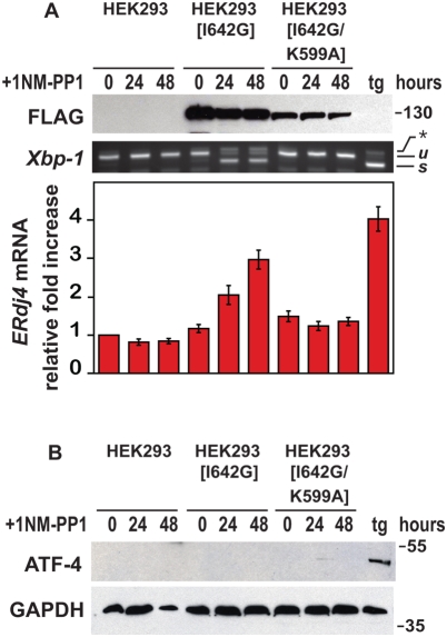
Figure 2. Selective and specific activation of IRE1 signaling.
(A) Parental wild-type and transgenic HEK293 cells expressing the Ire1[I642G], or Ire1[I642G/K599A] allele were treated for the indicated times with 1NM-PP1 (1 µM), and wild-type cells were treated for 4 hours with thapsigargin (tg) (300 nM). IRE1[I642G] and IRE1[I642G/K599A] protein was detected by immunoblotting for the FLAG epitope. GAPDH levels were assessed as a protein loading control. Xbp1 mRNA splicing was determined by RT-PCR. The unspliced (u) and spliced (s) Xbp1 mRNA products are indicated as labeled. ERdj4 mRNA levels were measured by quantitative PCR, normalized to Rpl19 mRNA levels, and are shown relative to levels in untreated cells. (B) Parental wild-type and transgenic HEK293 cells expressing the Ire1[I642G] or Ire1[I642G/K599A] alleles were treated for the indicated times with 1NM-PP1 (1 µM); wild-type cells were also treated for 4 hours with thapsigargin (tg) (300 nM). ATF4 protein was detected by immunoblotting. GAPDH levels were assessed as a protein loading control.

