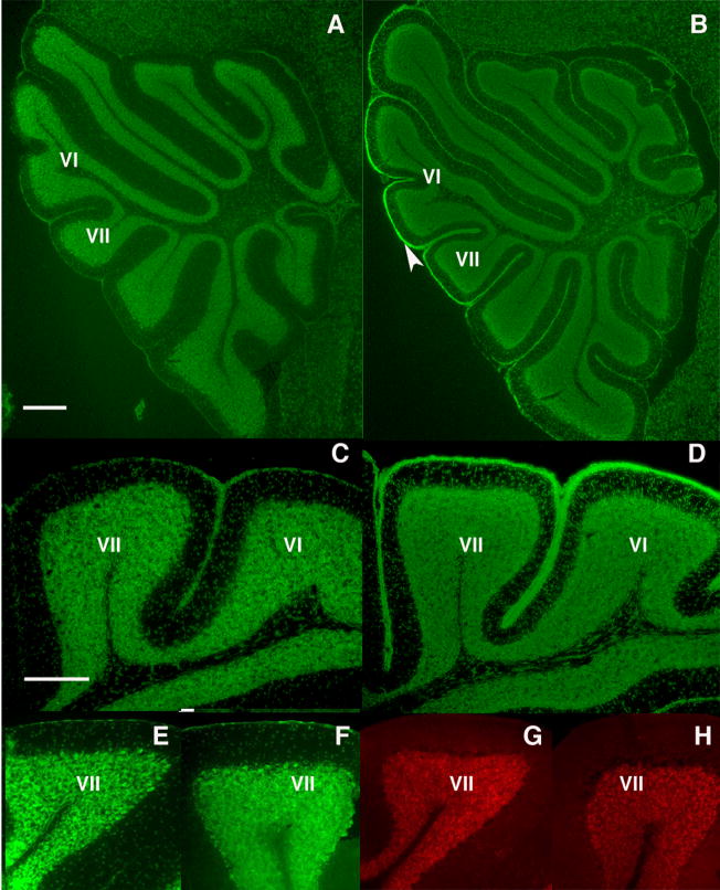
Figure 4.
Granule cell development is abnormal in the offspring of infected mothers. At P17, control mice lack an EGL (A, C), while a persistent EGL is observed in the offspring of infected mothers (B, D), particularly around lobules VI and VII (arrowhead). In the adult, Nissl staining reveals the normal absence of an EGL in both control (E) and experimental (F) offspring. Moreover, no GABAR α6 staining is found in the ML of the adult control (G) or experimental (H) offspring, indicating that the GCs have completed their migration into the IGL. Scale bars A, B = 200μm; C–H = 100μm.

