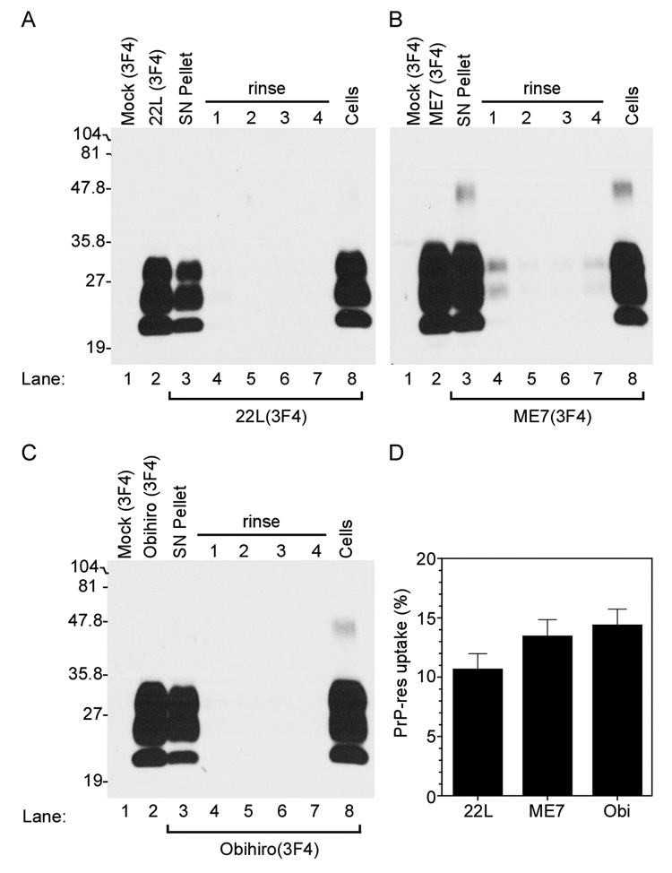
Figure 2. Mouse neural cells take up PrP-res3F4.
Western blot analysis of PrP-res3F4 uptake into MoL42-CFD5 cells. Brain homogenate from 22L, ME7 or Obihiro scrapie-infected Tg(WT-E1) mice were PK treated and used as positive controls (Panels A–C, lane 2) while PK treated brain homogenate from mock-infected Tg(WT-E1) mice was used as negative controls (Panel A–C, lane 1). For both the positive and negative controls, 1/20th of the total homogenate used was loaded onto the gel. Brain homogenates (200µl of a 1% brain homogenate) from 22L(3F4) (panel A), ME7(3F4) (panel B), or Obi(3F4) (panel C) were added to MoL42-CFD5 cells for 24 hours and then removed from the cell monolayer. Cell debris and dead cells were spun out of the supernatant and the supernatant pellet was PK treated, methanol precipitated and the entire sample loaded onto the gel (SN Pellet). Cells were then rinsed with PBS four times (rinse 1–4), lysed, and both the cell lysate and rinses were PK treated, methanol precipitated and the entire sample loaded onto the gel. PrP-res3F4 was detected in the lysed and PK treated cells (Cells). All blots were analyzed using the mouse monoclonal antibody 3F4 and developed using ECL (Amersham). The percentage of total PrP-res3F4 taken up by the cells after 24 hours is shown in Panel D. PrP-res3F4 was quantified as detailed in the materials and methods. There was no significant difference between the three strains in the amount of PrP-res taken up by the MoL42-CFD5 cells (p>0.05 using 1-way Anova with Bonferroni’s multiple comparison test). Data are shown as mean ± S.D. for N=12.

