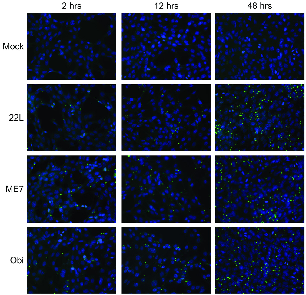
Figure 6. PrP-res3F4 uptake into neural cells using immunofluorescence microscopy.
PrP-res3F4 partially purified from 22L(3F4), ME7(3F4), Obi(3F4) or mock brain homogenates was added to MoL42-CFD5 cells for 2 – 48 hrs. Cells were rinsed, fixed and immunolabeled with the mouse monoclonal antibody 3F4. Anti-mouse FITC labeled antibody (green) was used to detect PrP-res3F4 while DAPI stain (blue) denotes cell nuclei. All images were taken with a 40X objective.

