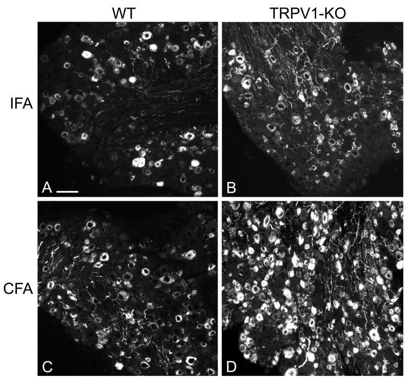
Figure 1.
Immunofluorescent staining for CGRP in DRG from sections of wild type (WT) and TRPV1-KO mice: the number of labeled neuronal profiles appears greater on the CFA-injected side than on the IFA-injected side 21 days after induction of AIA. In every group (WT and TRPV1-KO), the sections of are from the left and right L5 DRG of the same mouse. Scale bar, 100 μm.

