
Figure 2. Structure of the Ugene locus.
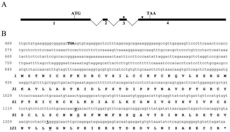
A) Numbered black boxes denote the 4 Ugene exons, with locations of initiator ATG and termination TAA designated. Sites which differentiate Ugene-p and Ugene-q are indicated by arrow heads. B) Nucleotide and deduced amino acid (aa) sequence of complete Ugene (Ugene-p) coding region. The Ugene nucleotide sequence is provided in the upper reading frame and numbered in roman type. The deduced aa sequence is provided underneath the nucleotide sequence, numbered in italic. The in-frame stop codon (TGA) 5′ of the start codon is indicated in boldface. Underlined letters represent codons that differ in Ugene-q.