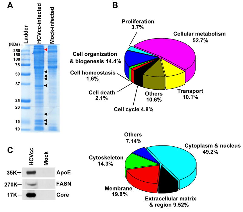
Figure 1. Identification of supernatant proteins that co-fractionated with HCV virions.
(A) Equal volumes of supernatant collected from HCVcc-infected and mock-infected Huh7.5.1 cells were subjected to filtration concentration and sucrose cushion ultracentrifugation. The pellets were dissolved in Laemmli buffer and resolved on a 10% one-dimensional SDS-PAGE gel. Eight representative protein bands differentially expressed between the samples are indicated with arrows and the red arrow points to the position of FASN. (B) Classification by gene ontology of the co-purified proteins with HCV virions in terms of their biological function and subcellular localization. (C) Protein pellets prepared from A were analyzed by western blotting and the presence of ApoE, FASN, and HCV Core protein was only found in the supernatant of HCVcc-infected cells.

