
Fig. 3.
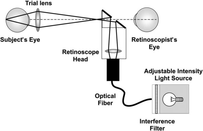
Schematic illustration of the chromatic retinoscope used to collect data from adults and infants, demonstrating the illumination path. The LCA of the lens in the retinoscope head changes the position of the secondary light source with wavelength. This change in position changes only the speed of the reflex motion rather than its direction and therefore does not influence the measurement of the eye's LCA. The LCA of the trial lenses used to assess the reflex was less than 0.1 D between 472 and 652 nm, for the lenses between +6 D and −5 D used to make the measurements. The radiant exposure levels for all wavelengths used in this experiment arriving at the cornea were also measured and confirmed to be at least 50 times lower than the appropriate ANSI safety standard when the light source was turned to its highest setting [37]. The LCA measurements were collected from subjects with the light source at the lowest setting that provided reliable data, and so the highest setting was never used during data collection.