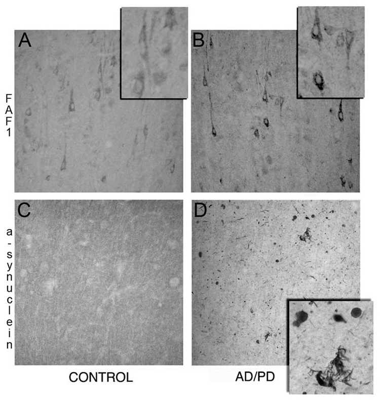
Fig 2. FAF1 expression in cortical tissue from AD/PD patients.
Paraformaldehyde fixed frontal cortex tissue from control (A, C) and AD/PD (B, D) patients were immunostained for FAF1 (A & B) and α-synuclein (C &D). FAF1 immunmoreactive cortical neurons can be identified in the control cortex. FAF1 expression can be observed in cell bodies and proximal dendrites while the nucleus appears to be devoid of FAF1 expression. Note increase in FAF1 expression in cortical neurons and neuropil and extensive α-synuclein pathology including Lewy bodies and neurites in AD/PD brain.

