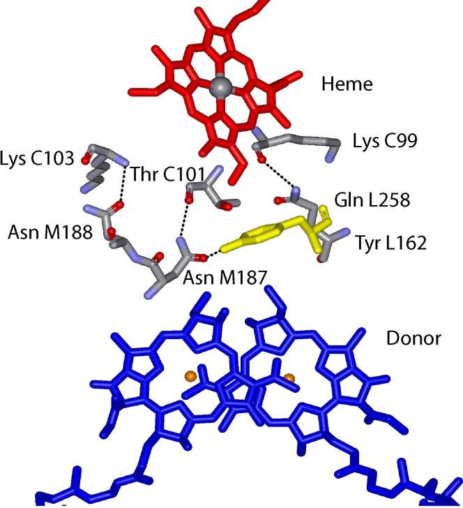
Figure 1.
Structure of the cyt c2:RC complex (PDB ID 1L9J) showing the inter-protein hydrogen bonds (dotted lines) from three amide residues on the RC to backbone atoms on the bound cyt c2. The redox active heme cofactor (red) on the cyt and the BChl2 donor (blue) on the RC, buried in their respective proteins, are in close contact with the key RC residue Tyr L162 (yellow) in the docked structure. (9)

