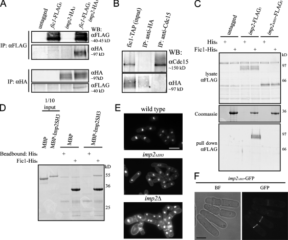
Figure 6.
Fic1 binds the SH3 domain of PCH family member Imp2. (A) Coimmunoprecipitations of Imp2-HA3 and Fic1-FLAG3 were performed as in Fig. 4 (B and C). IP, immunoprecipitation. (B) Fic1-TAP–containing complexes were purified from nda3-km311 imp2-HA3 cells using IgG beads followed by TEV cleavage. These complexes were immunoprecipitated using either an anti-HA antibody or anti-Cdc15 serum, and bound proteins were detected by immunoblotting. WB, Western blot. (C) Lysates from wild-type, imp2-FLAG3, or imp2ΔSH3-FLAG3 cells were incubated with recombinant bead-bound His6 or Fic1-His6. Bound proteins were detected by immunoblotting. (D) An in vitro binding experiment was performed with MBP-Imp2SH3 and Fic1-His6 as in Fig. 4 E. (E) Wild-type, imp2ΔSH3-HA3, and imp2Δ cells were fixed and imaged as in Fig. 1 A (quantitation in Fig. S4 D, available at http://www.jcb.org/cgi/content/full/jcb.200806044/DC1). (F) Imp2ΔSH3-GFP was visualized in live cells. BF, bright field. Bars: (E) 10 μm; (F) 5 μm.

