Figure 5.
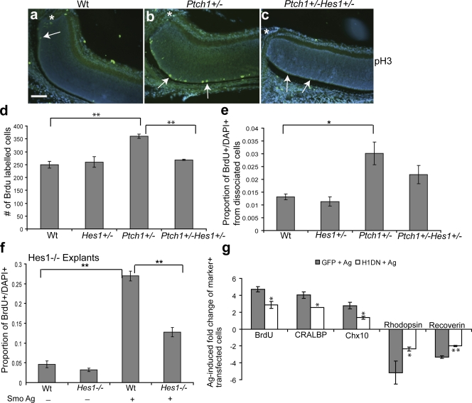
Shh-mediated RPC proliferation and cell fate specification requires Hes1. (a–c) In vivo anti-pH3 staining of the central retina adjacent to the optic nerve (asterisks) in P5 wild-type (Wt), PtchlacZ+/−, and PtchlacZ+/−Hes1+/− retinas. Arrows indicate pH3-positive cells. Note that pH3+ cells in the vicinity of the optic nerve are rare in Wt and compound heterozygous mice. Bar, 100 μm. (d) Quantitative analysis of BrdU incorporation in vivo from P5 Wt (n = 3), Hes1+/− (n = 3), PtchlacZ+/− (n = 3), and PtchlacZ+/−Hes1+/− (n = 6) retinas. Values represent the mean number of BrdU-positive cells counted from three sections per animal. (e) Quantification of the proportion of BrdU+ cells in single-cell dissociates from the retinas of Wt (n = 5), Hes1+/− (n = 3), PtchlacZ+/− (n = 8), and PtchlacZ+/−Hes1+/− (n = 7) retinas at P5. (f) Retinal explants from Hes1−/− (n = 3) or Wt (n = 3) animals were treated with a Smo agonist for 3 d, dissociated, and scored for the proportion of BrdU+DAPI+ cells. (g) Quantitative analysis for BrdU, CRALBP, Chx10, rhodopsin, and recoverin-positive cells in Smo agonist–treated P0 retinal explants electroporated with GFP and Hes1DN. Values are based on scoring marker+ cells among the transfected cohort in dissociates from retinal explants and represent the fold induction of double-positive (marker+GFP+) cells in GFP + Ag and Hes1DN + Ag cultures compared with double-positive cells in GFP-transfected untreated explants. There is no difference in proliferation or cell type composition in GFP and Hes1DN-transfected cells in untreated explants. Error bars represent SEM. *, P < 0.05; **, P < 0.01.

