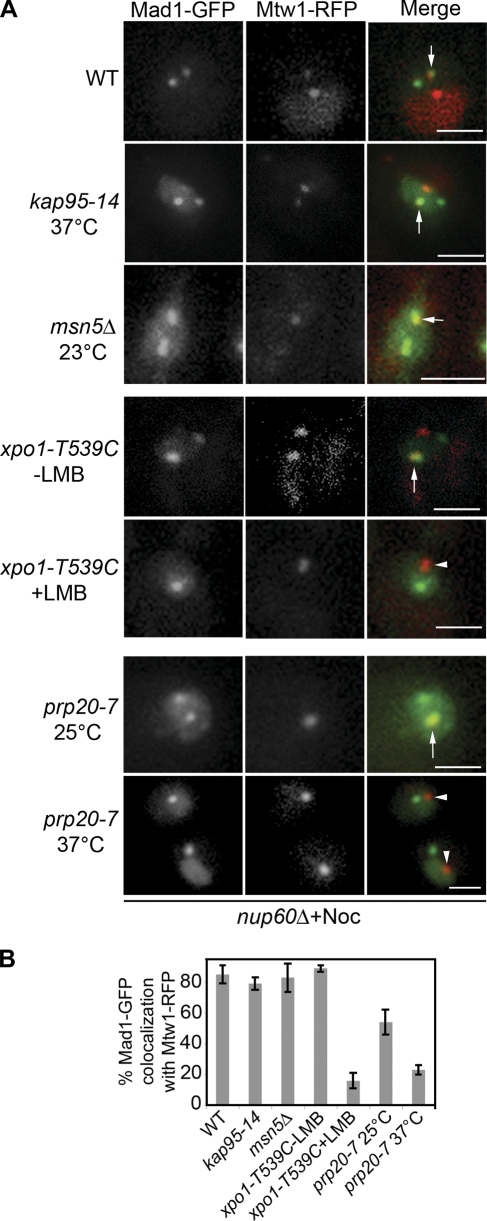
Figure 2.
Xpo1p-dependent targeting of Mad1p to kinetochores. (A) MAD1-GFP and MTW1-RFP in nup60Δ strains containing an otherwise WT genotype (Y3057) or the mutant alleles kap95-14 (Y3161), msn5Δ (Y3116), xpo1-T539C (Y3156), or prp20-7 (Y3091) were examined under the indicated conditions of temperature or LMB addition and after nocodazole addition. The localization of Mad1-GFP and the kinetochore protein Mtw1-RFP was examined by confocal microscopy. Note that as described previously (Gillett et al., 2004), some nocodazole-treated cells contain two kinetochore foci, with one lacking attached microtubules and being capable of binding Mad1p. Arrows point to overlapping Mad1-GFP and Mtw1-RFP foci. Arrowheads point to kinetochores devoid of Mad1-GFP. (B) Quantification of Mad1-GFP and Mtw1-RFP colocalization from the experiments presented in A. The results of three experiments were combined, and SD (error bars) is shown in each case. Bars, 2 μm.

