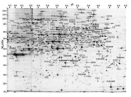
Figure 4.

2D PAGE yeast reference map with protein identifications as determined in the current investigation. The protein pattern corresponds to [35S]methionine-labeled polypeptide (19). The arrow indicates the protein sequenced in Fig. 3 and the rectangles indicate proteins of the yeast L-A virus. The elipses mark locations of very weakly staining proteins. Gene names of previously uncharacterized open reading frames are in boldface type.
