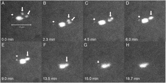
Figure 3. Achiasmate Xs can be visualized on the same half of the spindle in live oocytes.
Time-lapse of a live FM7/X oocyte is shown with only DNA fluorescence. A) Both achiasmate Xs (arrows) can be visualized on the right side of the spindle. In B and C the Xs move together and rejoin. In (D–H) the rejoined homologs move together back to the spindle midzone. Arrows point to separated or associated X chromosomes, while arrowheads point to separated or associated 4 th chromosomes, when they can be observed apart from the main chromosomal mass.

