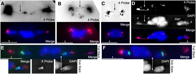
Figure 6. DAPI threads contain heterochromatin.
(A) An oocyte from FM7/X stained with DAPI (blue) and a FISH probe that preferentially labels chromosome 4 heterochromatin (red). When the FISH signal is greatly increased (top panel), a faint thread can be seen running between 4 th chromosomes, despite being separated by over 10 microns. (B) An oocyte from FM7/X stained with DAPI and 4 th probe. There is a clearly visible complete thread labeled with FISH probe running between 4 th chromosomes (arrow). (C) An oocyte from FM7/X stained with DAPI and 4 th probe that has reached the compact metaphase-arrested configuration. The 4 th probe reveals an incompletely-labeled thread running between the 4 th chromosomes, through the middle of the compact mass, suggesting that meiotic threads are not resolved until anaphase. (D) An oocyte from FM7/X stained with DAPI and 4 th probe, showing a clearly visible thread in both channels (arrows) that begins at the left 4 and appears to terminate at the adjacent X. (E) An oocyte from FM7/X stained with DAPI (blue), 4 th probe (red), and a probe that preferentially labels a block of the heterochromatin on the X (green). The FM7 chromosome (right, and insets) shows DAPI threads, but these threads do not appear to incorporate the X FISH probe. (F) An oocyte from FM7/X stained with DAPI, 4 th, and X FISH probes, showing a DAPI thread coming off the normal sequence X (right, and inset) that does not appear to incorporate the X FISH probe.

