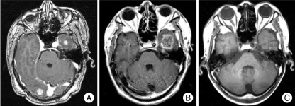
Fig. 3.
Serial magnetic resonance image (MRI) scans show the development of radiation necrosis and resolution. A : 66 year-old male patient was treated gamma knife radiosurgery (GKS) for 19 lesions with (radiation necrosis) marginal dose of 15 Gy at 50% isodose. B : Brain MRI at 7 months after GKS reveals increase in size of rim-enhancing lesion on left temporal lobe. C : The lesion was not treated furthermore and brain MRI at 38 months after initial GKS shows decrease in size and no enhancement of this lesion.

