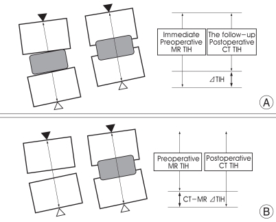
Fig. 2.
Diagram showing measurement of total inter-vertebral height (TIH) of the two fused vertebral bodies, the black arrow head is the mid-point of upper end plate of cranial vertebral body, the white arrow head is the mid-point of lower end plate of caudal vertebral body and the arrow line indicates TIH. A showing measurement of ΔTIH (difference between the immediate postoperative TIH and the follow-up TIH in the lateral radiographs). B showing measurement of computed tomography (CT)-magnetic resonance (MR) ΔTIH (difference between TIH of the postoperative mid-sagittal 2D CT and that of the preoperative mid-sagittal T1-weighted MR). The degree of subsidence was reflected by ΔTIH and the change of postoperative disc space height was reflected by CT-MR ΔTIH.

