Fig. 1.
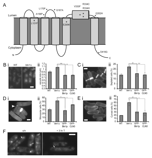
btn1Δ cells have multiple phenotypes which can be rescued by ectopic expression of Btn1p or CLN3.(A) Predicted topology of CLN3, based on Nugent et al., 2008. Missense mutations are marked with asterisks. (B) btn1Δ cells have larger vacuoles than wild-type cells: (i) vacuoles of wild-type (left panel) and btn1Δ (right panel) cells stained with FM4-64, (ii) bar chart of the mean vacuole size of the indicated cells. (C) btn1Δ cells have a higher septation index than wild-type cells: (i) btn1Δ cells stained with calcofluor to visualise cell walls and septa, (ii) bar chart of the percentage of the indicated cells with a visible septum. (D) btn1Δ cells have a defect in the initiation of bipolar growth after 7 hours at 37°C: (i) btn1Δ cells stained with calcofluor to show growing ends, the filled arrowhead indicates a monopolar cell with calcofluor staining absent at one pole, (ii) bar chart of septated cells with monopolar calcofluor staining. (E) btn1Δ cells are curved by 4 hours at 37°C: (i) btn1Δ cells stained with calcofluor, (ii) bar chart of the percentage of bent or curved cells. (F) Localisation of GFP-Btn1p in btn1Δ cells. Localisation of GFP-Btn1p after overnight expression (left panel) and 3 hours after promoter repression (right panel). Arrow=vacuole, filled arrowhead=endoplasmic reticulum (ER), unfilled arrowhead=pre-vacuolar compartment. (Data shown is the mean±s.d. of at least three independent experiments; ***P<0.001, **P<0.01, *P<0.05.) Bars, 5 μm.

