Fig. 4.
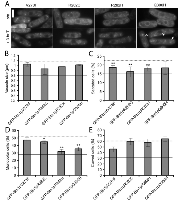
Mutations in the third lumenal loop, including the predicted amphipathic helix, impair most functions of Btn1p. (A) Localisation of GFP-tagged mutants at steady state (upper panels) and after promoter repression (lower panels). Arrow=vacuole, filled arrowhead=ER, unfilled arrowhead=pre-vacuolar compartment. (B-E) Phenotypes of btn1Δ cells expressing the indicated mutant GFP-Btn1p proteins: (B) mean vacuole diameter (μm); (C) mean septation index (% of total cells); (D) mean percentage of monopolar cells (% of total septated cells after 7 hours at 37°C); and (E) mean percentage of curved or bent cells (% of total cells after 4 hours at 37°C). Dotted line=mean value for btn1Δ cells, unbroken line=mean value for btn1Δ cells + GFP-Btn1p. (Data shown is the mean±s.d. of at least three independent experiments; ** P<0.01, *P<0.05.) Bar, 5 μm.

