
Fig. 9.
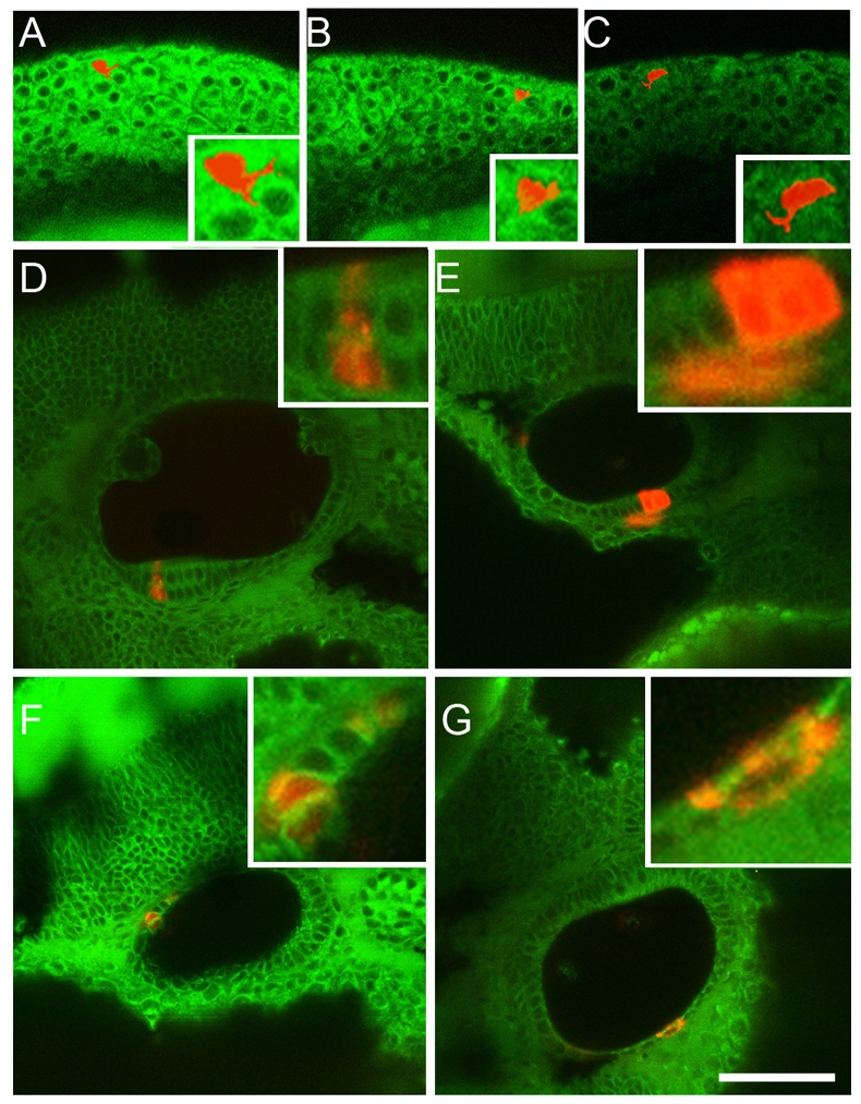
Neural crest cells make a minor, and transient, contribution to the otic vesicle. Single premigratory neural crest cells in the rhombomere 4 region of WT embryos that have been labelled iontophoretically with rhodamine dextran. The cells are shown at the 10-somite stage, approximately 1 hour after labelling (A–C, three different cells). The progeny of single labelled cells that contribute to the otic epithelium are shown at 36 hpf, approximately 24 hours after labelling (D–G, four different examples). The insets show closeups of each cell or clone. Note that occasional progeny incorporated into the otic epithelium include cells in the sensory epithelium of the anterior macula (D). The clone in (E) includes three cells in the otic epithelium, plus one lying outside the epithelium. The fate of this latter cell is not known. Bar, 25 μm.