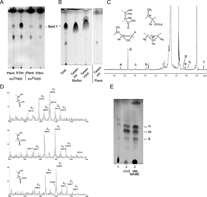
Fig. 4.

M. tuberculosis biofilms contain free mycolic acids.
A. 1D-TLC of apolar lipids extracted from either M. tuberculosis mc27000 cells or the mc27025 mutant growing either planktonically or as biofilms resolved in solvent system C. Samples were labelled with 14C-acetate and equal amounts of total radioactivity were examined.
B. Tween-80 solubilization of biofilm lipids. Biofilm samples were Tween-80 extracted and the Tween-soluble and Tween-insoluble fractions separated by TLC as above. The Tween-containing supernatant from planktonically grown cells was analysed similarly.
C. 1H-NMR spectrum of Spot 1. 1H signals of protons associated with functional groups were assigned using known chemical shifts (Watanabe et al., 1999; X. Trivelli and Y. Guerardel, unpubl. obs.) and labelled (a to h) according to their relative positions. A slight de-shielding of Ha (Δδ + 0.06 p.p.m.) proton from 3.64 to 3.70 p.p.m. and Hb (Δδ + 0.03 p.p.m.) proton from 2.43 to 2.46 p.p.m. compared with their equivalent protons associated to a methyl-esterified carboxyl group is in agreement with the occurrence of a free mycolate.
D. MALDI-TOF-MS analysis of native (top panel), -CH3 (middle panel) and -CD3 (bottom panel) esterified Spot 1. Signals correspond to [M + Na]+ adducts of a family of methoxy mycolates.
E. Spot 1 (lane 1) was methyl-esterified (lane 2) and compared with methyl-esterified mycolic acids purified from Mtb cell wall (lane 3), by resolution on a TLC plate developed in petroleum ether : acetone (95:5) and visualized as in Fig. 4A. a, m and k indicate the alpha, methoxy and keto mycolic acids respectively.
