
Fig. 5.
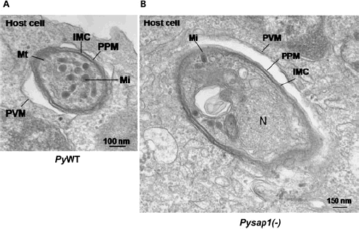
Pysap1(−) liver stages form a parasitophorous vacuole membrane (PVM). Electron microscopic analysis establishes that intrahepatocytic Pysap1(−) parasites form a PVM.
A. Transversal section of a PyWT parasite within a HepG2-CD81 cell 1 h post infection. The PVM is indicated.
B. Transversal section of Pysap1(−) sporozoite 1 h post infection in HepG2-CD81. The PVM is indicated. IMC, inner membrane complex; Mi, microneme; N, nucleus; PPM, parasite plasma membrane; PVM, parasitophorous vacuole membrane; Mt, mitochondrion.