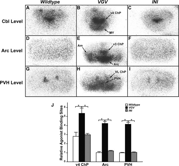
Figure 10.

Increased binding of 5-HT2CR-selective radioligands in VGV mouse brains. A–I, The brain structure was visualized by binding of (±)-[125I]DOI agonist to 5-HT2C receptors. Representative autoradiographs are shown. 5-HT2C receptors were visualized with 5-HT2A/2CR agonist (±)-[125I]DOI (0.14 nm) in the presence of 10−7 m cold spiperone, the 5-HT2AR antagonist. PVH, Paraventricular nucleus of the hypothalamus; Arc, arcuate nucleus of the hypothalamus; Cbl, Cerebellum; Am, amygdala; ChP, choroid plexus; v3, third ventricle; v4, fourth ventricle; VL, lateral ventricle; MY, medulla. J, Quantification of 5-HT2CR agonist binding sites were done by measuring the optical density of areas representing the fourth ventricle choroid plexus (v4ChP), Arc, and PVH (n = 3). The relative optical density normalized to the density determined for the PVH area of the wild-type mouse brain is indicated. Significant differences are indicated by asterisks (*p < 0.05). Error bars indicate SEM.
