
Figure 1.
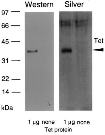
Electrophoretic characterization of TetA(L)–H6 purified from E. coli expressing a modified B. subtilis tetA(L) gene on a multicopy plasmid. Purified TetA(L)–H6 (1 μg) next to empty lanes containing no protein. After electrophoresis, the sample was silver-stained (Right) or transferred and analyzed for cross-reaction with antibody raised against a synthetic peptide corresponding to the N-terminal 14 amino acids of TetA(L) (Left); although not shown, preimmune rabbit serum showed no cross-reaction. The arrowhead indicates the position of TetA(L)–H6. A higher set of bands found in both lanes of the silver-stained gel is most likely a staining artifact.