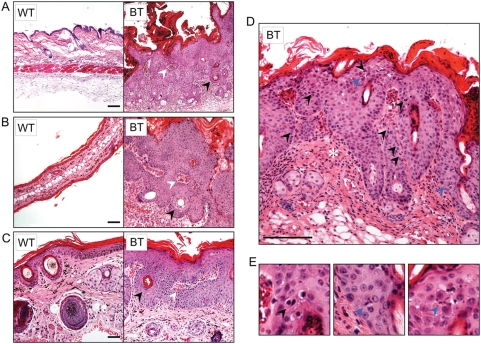
Figure 3. Ets1 expression causes hyperplastic and dysplastic changes in the skin epidermis.
Microscopic features of the cutaneous changes in (A) dorsal trunk, (B) ear and (C) tail skin of BT mice upon Ets1 induction show epidermal and sebaceous hyperplasia, hyperkeratosis, and parakeratosis, keratin pearls (black arrowheads), vascularization, leukocytic infiltration and dermal enclosures within the epidermal downgrowths (white arrowheads). (D) High power view of the dorsal epidermis demonstrating the presence of several mitotic figures including some in the suprabasal layers (black arrowheads). Reactive leukocytic infiltration is also evident (asterisk). (E) Further magnification of supra-basal mitoses in lesions of the dorsal skin in BT mice. Some of the mitoses exhibit abnormal, tripolar mitotic spindles (blue arrowheads).

