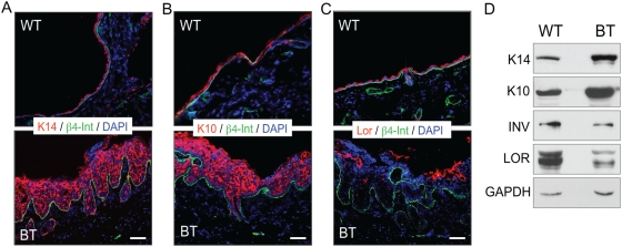
Figure 4. Ets1 expression disrupts the normal differentiation pattern of the skin epidermis.
Immunofluorescent staining of epidermal differentiation markers on dorsal skin of induced Ets1 BT mice and littermate wild type mice. Each section was stained with antibodies to β4- integrin to mark the position of the basement membrane and with DAPI to detect nuclei. Epidermal differentiation was assessed by staining for (A) the basal layer marker keratin 14 (K14), (B) the spinous layer marker keratin 10 (K10) and (C) the granular layer marker loricrin (Lor). (D) Western blot analysis of dorsal skin extracts to confirm results obtained in immunofluorescence. GAPDH serves as a loading control for Western blots.

