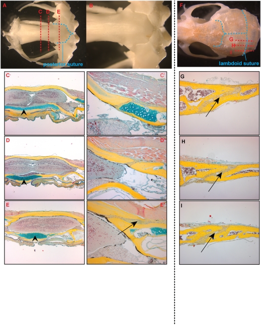
Figure 6. Morphology of the posterior suture.
A. Dry skull preparation of adult Xenopus demonstrating the frontonasal and posterior sutures (outlined in blue dashed lines). B. Dorsal view of posterior suture at 1.25× magnification. C–E. Pentachrome stained coronal sections of an adult skull (5× magnification). The location of the planes of section are notated by red dashed lines in the dry skull prep in A. C′–E′. 20× magnification of suture. There are no midline sutures evident but at the posterior aspect, lateral overlapping sutures are present (Arrow in E′ note overlapping pattern). The skull base ventral to the brain has a cartilaginous layer throughout (arrowheads). F. Dry skull preparation of adult mouse demonstrating the lambdoid suture. G–I. Pentachrome stained parasagittal sections of adult mouse calvaria. Note the overlapping pattern of the suture (arrows).

