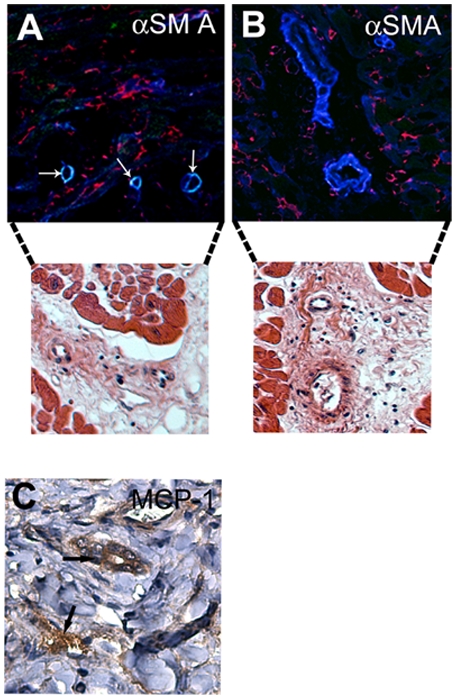
Figure 4. Staining of mouse cardiac allografts for αSMA to identify the origin of cells expressing αSMA.

(A and B) Host-derived SMCs were present in arterioles with a single layer of SMCs (yellow) (A) but not in those with more SMC layers (B). Arrows indicate host-derived cells. Blue, αSMA; green, green fluorescent protein; red, nuclear counterstaining. Confocal microscopy analysis is followed by hematoxylin-eosin staining of parallel sections in order to present structure of vessels. Staining of human cardiac allografts for MCP-1 revealed MCP-1 around the small arterioles in an area with inflammation (C).
