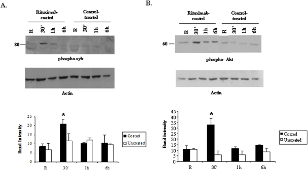
Figure 2. Rituximab-coated targets induce significant activation of Syk kinase and the PtdIns 3-kinase/Akt pathway in macrophages.
IFNγ-primed peritoneal macrophages were stimulated with paraformaldehyde-fixed Rituximab-coated or control antibody-treated Raji cells at E∶T ratio of 1∶1 for indicated time points. Protein-matched whole cell lysates were analyzed by Western blotting with A. phospho-Syk antibody (upper) and B. phospho-Akt antibody (upper). The same membranes were reprobed with actin antibody (lower). The graphs in the lower panels show mean and SD of the band intensity of phospho-Syk and phospho-Akt from three independent experiments. Data were analyzed by student's t-test (* = p value≤0.05).

