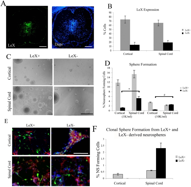
Figure 4. LeX negative cells from spinal cord, but not cortical derived neurospheres are neural stem cells.
(A) LeX expression in a coronal section of the embryonic day 15 spinal cord; LeX (Green) Dapi (Blue); Dorsal (top) ventral (bottom). (B) Percentage of cells in secondary neurospheres derived from cortex and spinal cord that express LeX. (C) Photomicrographs of spheres generated from E14 cells following sorting for LeX expression (at second passage). LeX negative cells from cortical derived neurospheres tend to form clusters rather than round phase bright neurospheres. (D) Quantification of neurosphere formation from E14 cortical and spinal cord derived cells following sorting. Sorted cells were cultured at clonal density (1,000 cells/ml) and 10,000 cells/ml. (E) Immunocytochemistry of differentiated E14 clonal neurospheres generated by LeX expressing and non expressing cells from cortical and spinal cord derived neurospheres. Upon differentiation, clusters generated by LeX negative cells from cortical neurospheres lose cell integrity and do not generate morphologically distinct cell types. TuJ1 (Green), O4 (Blue), GFAP (Red). (F) Ability of cells within a sphere generated by LeX+ and LeX− cells to form a new neurosphere. Bars are mean±SEM of at least 3 independent experiments. * P<0.001, # P<0.0000005, Anova followed by post hoc t-test. Scale bar in A: 450 µm D: 200 µm and in E: 110 µm.

