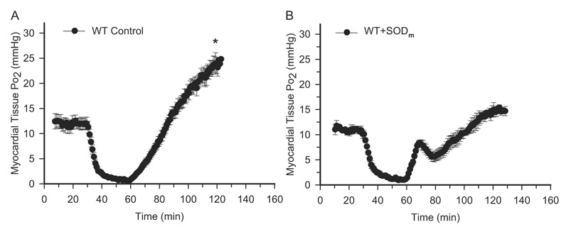
Fig. 2.
EPR oximetry on WT control and SODm-treated mouse hearts. After thoracotomy and exposure of the heart, ~10 µg of LiPc was implanted into the midmyocardium in the risk area, and EPR spectra were recorded. Time-dependent EPR line width was measured, and tissue Po2 was calculated. A, myocardial tissue Po2 before and during ischemia and after reperfusion in WT control mouse hearts. B, myocardial tissue Po2 before and during ischemia and after reperfusion in SODm-treated mouse hearts (bolus intravenous injection at a dose of 4 mg/kg 5 min before reperfusion). These measurements demonstrated that there is an overshoot of tissue Po2 after reperfusion, and this hyperoxygenation is attenuated after treatment with SODm. In addition, there is a transient peak of tissue Po2 at 9 min of reperfusion in SODm-treated mouse hearts. *, p < 0.01, WT Control versus WT + SODm at 60-min reperfusion, n = 7.

