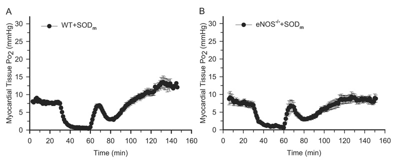
Fig. 3.
EPR oximetry on WT SODm-treated and eNOS−/− SODm-treated mouse hearts. Surgical procedures and EPR measurements were the same as described in Fig. 1. SODm was given as a bolus (4 mg/kg) at 30 min before ischemia. A, myocardial tissue Po2 before and during ischemia and after reperfusion on WT SODm-treated mouse hearts. There is a transient peak of tissue Po2 at 9 min after reperfusion, even when SODm was given 30 min before ischemia. B, myocardial tissue Po2 before and during ischemia and after reperfusion on eNOS−/− SODm-treated mouse hearts. The transient peak of Po2 exists even in the eNOS−/− mouse hearts. n = 7 for each group. These measurements demonstrated that the transient peak of tissue Po2 after treatment with SODm is independent of endothelium-derived NO.

