
Figure 2. Reprogramming of non-terminally differentiated B cells to pluripotency.
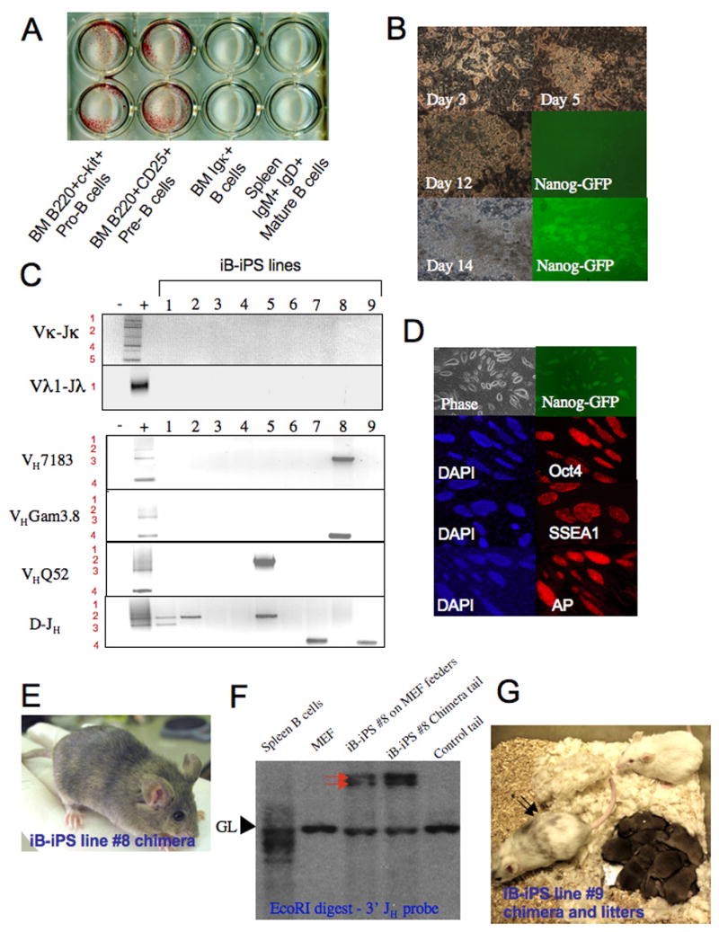
(A) 8 week old chimera derived bone marrow B cell subsets (50*10^3 cells per well) and spleen IgM+IgD+ mature B cells (250*10^3 spleen cells per well) were plated on OP9 bone marrow stroma with conditioned media and Dox. After 14 days plates were fixed and stained for AP activity. (B) Images for characteristic colonies at different time point obtained from sorted B220+CD25+ bone marrow cells. (C) PCR analysis for rearrangements in selected iB-iPS. MEFs and splenocytes were used as negative and positive controls, respectively. (D) Immunostaining for ES cell markers on a representative line iB-iPS#8. (E) A chimeric mouse from iB-iPS #8 cell line. Agouti colored hairs originate from injected iPS lines. (F) Southern blot analysis of iB-iPS line #8 grown on MEF feeders and of tail tip biopsy taken from the derived chimera. MEF and spleen B cells were used as negative and positive controls, respectively, for rearrangement detection. Red arrows indicate the two rearranged heavy chain alleles. GL indicates germline fragment representing non-rearranged configuration of the Igh locus. (G) An adult old chimeric pseudo-male mouse from iB-iPS #9 cell line was mated with Balb/C females and repeatedly achieved 100% germline transmission, as indicated by the agouti color of all litters obtained.