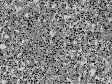
Fig. 6.
The parathyroid adenoma was composed of a single cell proliferation of enlarged chief cells without any fat cells present. The chief cells had clear to slightly eosinophilic cytoplasm and round to oval nuclei with heavy chromatin distribution. The cells are arranged in a glandular and solid pattern (Hematoxlyin-eosin, × 200).

