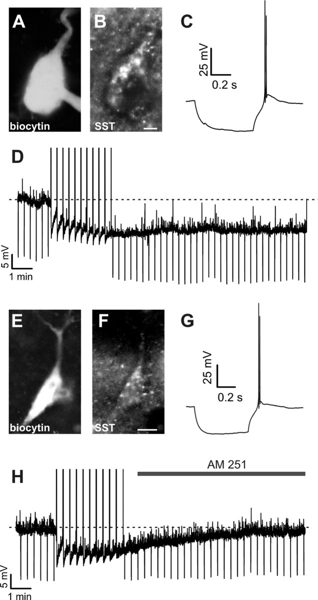
Figure 1.

CB1-dependent SSI in neocortical LTS interneurons expressing the neuropeptide somatostatin. A, Example of a biocytin filled interneuron, processed with Cy3-conjugated streptavidin. B, The same slice was counterstained using an antibody against the neuropeptide SST, indicating that the same cell was SST-positive. C, Firing properties of the cell of A-B, showing the typical rebound burst overriding a low-threshold spike (LTS) in response to a hyperpolarizing current step (−70 pA). Vm was −62 mV. D, The same cell as in A–C showed a persistent hyperpolarization (slow self-inhibition, SSI) in response to 10 trains of action potentials at 50 Hz (intertrain intervals: 20 s). Negative deflections represent responses to negative current injections (−30 pA), showing reduced gm associated to the hyperpolarization. E, Example of another biocytin filled interneuron processed with Cy3-conjugated streptavidin. F, The cell of E showed SST-immunoreactivity. G, The cell of E and F showed typical rebound LTS firing behavior in response to hyperpolarizing current injection as in C. Vm: −58 mV. H, Cell of E-G responded to SSI-inducing stimuli with a hyperpolarization, which was reversed by extracellular application of the CB1R antagonist AM 251 (3 μm). Scale bars: A, B, 2.5 μm; E, F, 5 μm.
