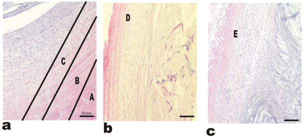
Figure 11.
Hemotoxylin and Eosin stains for the IVD-AF regions. Hematoxylin and Eosin stains bind to charged particles with in the tissue. The staining pattern of H and E resembles the pattern of Saf-O and Fast-G. The tissue appears to be more acidic in the inner regions compared to the outer regions likely due to the limited blood supply and hypoxic conditions of the inner AF. Image (a) shows A, B and C regions, while images (b) and (c) represent the regions D and E respectively.

